38 heart diagram with labels and blood flow
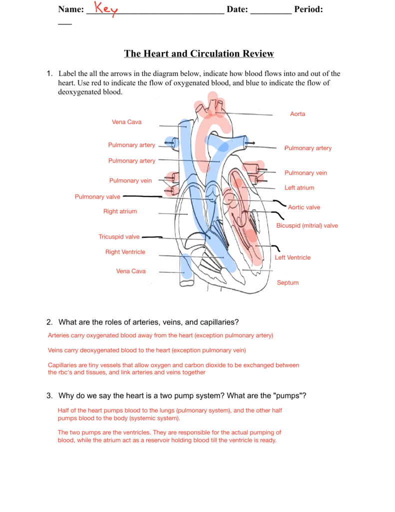
PDF Anatomy of Heart Labeled and Unlabeled Images (a) Anterior view of the external heart C' 2019 Pearson Education. Aort'c arch Ligamentum arteriosum Left pulmonary artery Left pulmonary ve ns Auricle of left atrium Circumflex artery Left coronary artery (in atrioventricular sulcus) Great cardiac vein Left ventricle Anterior interventricular artery (in anterior interventricular sulcus) Apex Heart Anatomy: Labeled Diagram, Structures, Blood Flow ... There are 4 chambers, labeled 1-4 on the diagram below. To help simplify things, we can convert the heart into a square. We will then divide that square into 4 different boxes which will represent the 4 chambers of the heart. The boxes are numbered to correlate with the labeled chambers on the cartoon diagram.
Diagram of Blood Flow in the Heart, Lungs and Body ... This medical illustration depicts a diagram of blood flow through the body. Oxygenated (oxygen-rich)blood travels from the lungs to the heart, where it is then pumped throughout the body. Deoxygenated (oxygen-poor) blood travels from the body back to the heart, where it is pumped to the lungs for gas exchange. Labels include the common carotid arteries, jugular veins, superior vena cava ...

Heart diagram with labels and blood flow
Heart Blood Flow Circulation Anatomical Diagram with ... Heart blood flow circulation anatomical diagram with atrium and ventricle system. Vector illustration labeled medical poster.. Illustration about cardiac, coronary, cardiovascular - 116079724 Heart Blood Flow | Simple Anatomy Diagram, Cardiac ... Step 1 and 6 involve a blood vessel, which makes sense as this is how blood enters and exits that side of the heart. Steps 2-5 involve a chamber, valve, chamber, and valve. So if you remember this general pattern, it will help you recall the order in which blood flows through each side of the heart. Right Side of the Heart SVC/IVC Right Atrium Cardiology: Basic Physiology Of The Heart And Mechanisms ... Arrows represent the flow of blood. Labels include the right and left common carotid arteries, pulmonary arteries, pulmonary veins, and lungs and the right and left atrium and ventricles, the left subclavian artery, aorta, jugular veins, thoracic aorta, inferior and superior vena cava and capillaries.
Heart diagram with labels and blood flow. Heart blood flow circulation anatomical diagram with ... Description: Heart blood flow anatomical diagram with atrium and ventricle system. Vector illustration labeled medical poster. Blood circulation path scheme with arrows. You may also like… Coronary circulation anatomical cross section diagram, labeled vector illustration scheme € 7.99 Add to cart Human Heart Diagram Stock Photos, Pictures & Royalty-Free ... Blood Flow Human Heart Cross Section of Heart with Labels on White Background circulatory system anatomy Antique illustration: heart diagram Heart failure or congestive heart failure Human Heart Heart blood flow circulation anatomical diagram with atrium and... Electrocardiogram and heart pattern background (health concept) A Diagram of the Heart and Its Functioning Explained in ... The heart blood flow diagram (flowchart) given below will help you to understand the pathway of blood through the heart.Initial five points denotes impure or deoxygenated blood and the last five points denotes pure or oxygenated blood. 1.Different Parts of the Body ↓ 2.Major Veins ↓ 3.Right Atrium ↓ 4.Right Ventricle ↓ 5.Pulmonary Artery ↓ 6.Lungs File:Heart diagram blood flow en.svg - Wikipedia File:Heart diagram blood flow en.svg. Size of this PNG preview of this SVG file: 330 × 370 pixels. Other resolutions: 214 × 240 pixels | 428 × 480 pixels | 535 × 600 pixels | 685 × 768 pixels | 913 × 1,024 pixels | 1,827 × 2,048 pixels. This is a file from the Wikimedia Commons. Information from its description page there is shown below.
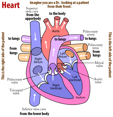
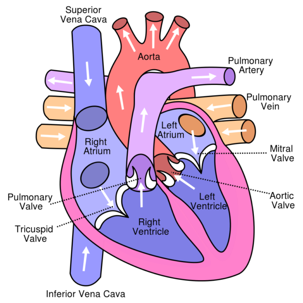
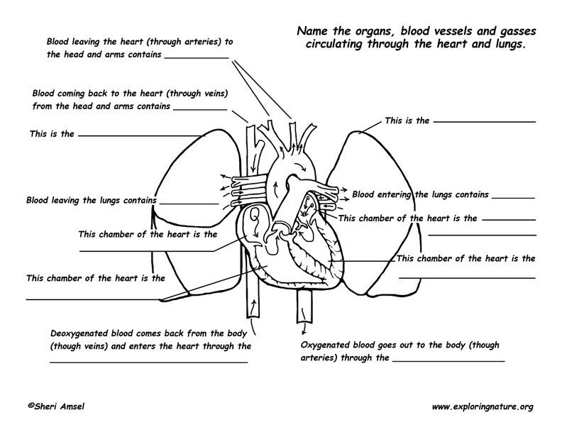
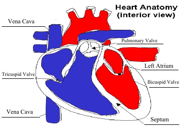
Diagram of Blood Flow Through the Heart - Bodytomy The heart is divided into two chambers, left and right, the right atrium and ventricle lie on the right side and the left atrium and ventricle on the left side. These two chambers are not directly connected to each other. Synchronization of the Two Chamber The right and left side or chambers of the heart work in tandem with each other. Diagram of Human Heart and Blood Circulation in It | New ... Every heart diagram labeledwill clearly show these valves. These valves allow blood flow in one direction only. Different valves perform different functions. Tricuspid valve is located between the right ventricle of your heart and the right atrium, and allows the blood to move from the right atrium to the right ventricle. Heart Diagram Flow Teaching Resources | Teachers Pay Teachers Cardiovascular System: Heart Diagram to Color by Lori Maldonado 80 $2.00 PDF This diagram shows the way blood flows through the heart. The areas of the heart with MORE oxygen are labeled with an "R". Students will color these areas RED. The areas of the heart with LESS oxygen are labeled with a "B". Students will color these areas BLUE. Circulatory System Diagram | New Health Advisor There are different types of circulatory system diagrams; some have labels while others don't. The color blue stands for deoxygenated blood while red stands for blood which is oxygenated. Below you'll see diagram specified to the heart, as well as circulatory system diagram of the whole body: How Does the Human Circulatory System Work? 1. Heart
Box Diagram, Labels of Heart, and Blood Flow through Heart ... About Press Copyright Contact us Creators Advertise Developers Terms Privacy Policy & Safety How YouTube works Test new features Press Copyright Contact us Creators ... Heart Diagram with Labels and Detailed Explanation The diagram of heart is beneficial for Class 10 and 12 and is frequently asked in the examinations. A detailed explanation of the heart along with a well-labelled diagram is given for reference. Well-Labelled Diagram of Heart The heart is made up of four chambers: The upper two chambers of the heart are called auricles. Label the heart — Science Learning Hub In this interactive, you can label parts of the human heart. Drag and drop the text labels onto the boxes next to the diagram. Selecting or hovering over a box will highlight each area in the diagram. Pulmonary vein Right atrium Semilunar valve Left ventricle Vena cava Right ventricle Pulmonary artery Aorta Left atrium Download Exercise Tweet Circulatory System Diagram - Cardiovascular System and ... SmartDraw has a number of templates included for circulatory system diagrams, cardiovascular system diagrams, blood circulation diagrams, and more. You don't really have to "draw" them as much as find them and modify them as needed. You can add labels or titles and change the size of symbols as necessary.
Cardiovascular system review key
Blood Flow Through the Heart - Austin Community College ... Blood Flow Through the Heart. Beginning with the superior and inferior vena cavae and the coronary sinus, the flowchart below summarizes the flow of blood through the heart, including all arteries, veins, and valves that are passed along the way. 1. Superior and inferior vena cavae and the coronary sinus 2. Rt. atrium 3.

Human Heart Diagram Without Labels | Human heart diagram, Heart diagram, Human heart
Human Heart Diagram - Side View and Top View It's pretty amazing to watch blood flow through the heart. To see how blood flows through the heart using an animation, please click here. So you know, the diagram above is showing a side view of the heart. You can clearly see the left and right atrium as well as the left and right ventricles. If you look closely, you can also see the aortic ...

Detailed Labeled Anatomy Human Body | jpg: labeled heart flow | Nursing | Pinterest | Heart ...
Circulatory System: Blood Flow Pathway Through the Heart ... Pathway of Blood Through the Heart. In this educational lesson, we learn about the blood flow order through the human heart in 14 easy steps, from the superior and inferior vena cava to the atria and ventricles. Come also learn with us the heart's anatomy, including where deoxygenated and oxygenated blood flow, in the superior vena cava, inferior vena cava, atrium, ventricle, aorta ...
Tips for How to Study the Cardiovascular System
YR 8 Topic 2 Circulatory System - AMAZING WORLD OF SCIENCE ... ABOUT THIS ACTIVITY: Illustrates the pathway of blood through the heart. The areas of the heart with MORE oxygen are labeled with an "R". Students will color these areas RED. The areas of the heart with LESS oxygen are labeled with a "B". Students will color these areas BLUE.
13+ Heart Diagram Templates – Sample, Example, Format Download | Free & Premium Templates
Human Heart Diagram Labeled | Science Trends Let's examine the anatomy of the heart along with some diagrams that show how the heart operates. Anatomy Of The Heart The human heart usually weighs somewhere between 10 to 12 ounces in men and between 8 to 10 ounces in women, and in terms of size is roughly the size of the fist.

Pin on Nursing School
Heart Diagram | Free Heart Diagram Templates Do you know that human heart system can be even more powerful than an electronic equipment? Wanna figure out why? Just refer to this originally designed Edraw heart diagram science template for more details.
Pulmonary Circulation - Through Heart and Lungs (Advanced*)
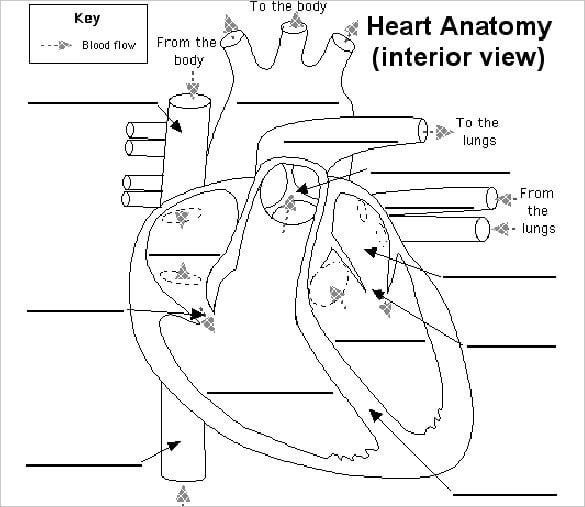
Heart Diagrams for Labeling and Coloring, With Reference ... (Not for black and white photocopying). - One black and white heart diagram with lines for students to fill in labels, and arrows showing blood flow - One black and white heart diagram with no lines or labels, but arrows included, so you can customize what labels the diagram will include
Cardiovascular System
Heart Anatomy & Circulatory System Blood Flow - Human ... Blood Flow Pathway of Blood Flow. Now that you understand how the heart and the circulatory system are organized, we can examine the pathway of blood in closer detail. Figure 5. Two labeled diagrams presenting the anatomy of the heart and the circulatory systems.

Heart Education - Pediatric Heart Specialists
Heart Diagram - 15+ Free Printable Word, Excel, EPS, PSD ... Heart Diagram - 15+ Free Printable Word, Excel, EPS, PSD Template Download A heart diagram is a popular design used by different people for various uses. It can be used by a teacher or student for academic purpose, by a friend or relative for mutually sending and exchanging cards or for baby toys or printing on dresses etc.

biology | Forgotten Physiology
PDF BLOOD FLOW THROUGH THE HEART diagram BLOOD FLOW THROUGH THE HEART diagram BE ABLE TO EXPLAIN THE BLOOD FLOW THROUGH THE HEART: RIGHT SIDE (REMEMBER ANATOMICAL POSITION) LEFT SIDE DEOXYGENATED BLOOD FROM BODY TISSUE OXYGENATED BLOOD FROM LUNGS VIA PULMONARY VEINS SUPERIOR AND INFERIOR VENA CAVA LEFT ATRIUM RIGHT ATRIUM BICUSPID VALVE

DIAGRAMS: Human heart blood flow diagram | nice post | Cardiac nursing, Medical assistant, Nurse ...
Cardiology: Basic Physiology Of The Heart And Mechanisms ... Arrows represent the flow of blood. Labels include the right and left common carotid arteries, pulmonary arteries, pulmonary veins, and lungs and the right and left atrium and ventricles, the left subclavian artery, aorta, jugular veins, thoracic aorta, inferior and superior vena cava and capillaries.

Diagram of a Heart Labeled and Unlabeled 2018
Heart Blood Flow | Simple Anatomy Diagram, Cardiac ... Step 1 and 6 involve a blood vessel, which makes sense as this is how blood enters and exits that side of the heart. Steps 2-5 involve a chamber, valve, chamber, and valve. So if you remember this general pattern, it will help you recall the order in which blood flows through each side of the heart. Right Side of the Heart SVC/IVC Right Atrium

Printable Heart Diagram
Heart Blood Flow Circulation Anatomical Diagram with ... Heart blood flow circulation anatomical diagram with atrium and ventricle system. Vector illustration labeled medical poster.. Illustration about cardiac, coronary, cardiovascular - 116079724
Heart Diagram Blood Flow Labeled
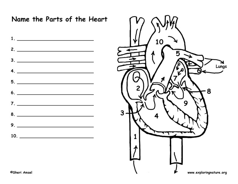
Heart and Blood Flow Labeling Page

Diagram of Heart Blood Flow for Cardiac Nursing Students | Cardiac nursing, Heart blood flow ...
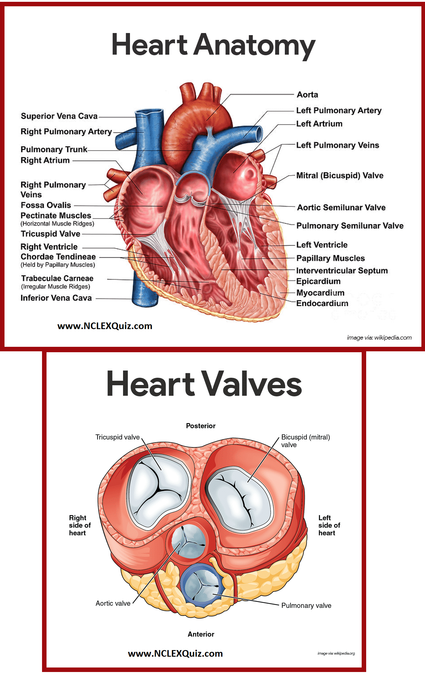
Diagram of Heart Blood Flow for Cardiac Nursing Students - NCLEX Quiz
Post a Comment for "38 heart diagram with labels and blood flow"